Liposomal Encapsulation Technology (LET) is an exciting new process that may revolutionize many forms of oral pharmacological therapy and more importantly may be in the realm of the common man to utilize. LET can offer a unique safe way to treat racehorses in a track situation without the use of banned syringes or needles. It is simply an encasing of small volumes of pharmaceutical compounds (herbs, too) by a phospholipid membrane (liposome) allowing safe passage through the gastrointestinal tract without degradation-directly into the blood plasma. It would be the closest thing we have to giving a pill that mimics an intravenous injection! Plus, liposmes could more easily assault and penetrate cell walls and many forms of infective biofilms resulting in a very effective delivery system against these pathological forms. As an example, it has been shown that 5 grams of Vitamin C which has been encased by liposomes is equal in results to 50 grams of Vitamin C that has been given intravenously! It appears that liposomal Vitamin C is far more efficient at permeating tissues than an IV application of Vitamin C. Vitamin C is mostly synthezied in the liver (mammals) or kidneys (reptiles and birds), so it makes sense that if a liposomal Vitamin C vehicle can reach the liver intact where the ascorbic acid is released and the phospholipid liposome is absorbed that it would be a very effective form of protocol. One can go out and purchase expensive Liposomal Vit C and other compounds, but it can be made at home very cheaply. Certainly the home-made Liposomal medications will not be quite as efficiently made as by the big med laboratories, but even if one can produce just 50% liposomal encapsulated material in the kitchen, that is more than good enough for our needs and can offer outstanding health benefits! My book, A Racehorse Herbal will study in detail new and exciting breakthroughs like LET which will more effectively treat the performance horse and will be within the capability of any horseman to process himself!

     Liposomes can be formed mechanically in three ways: 1) extrusion, 2) sonification, 3) microfluridization. The extrusion process forces the raw materials through some type of membrane. The sonification method is what we will be delving into and uses sound waves to stimulate liposomal formation. In this instance, we will use a common ultrasonic cleaner that can be bought just about any where. Lastly, microfluridization is the process the commercial companies seem to prefer and involves expensive equipment. It is more efficient than sonification. They use a microfluidizer to force the raw materials against a forming plate at extremely high pressures to form liposomes. The best liposomes are under 200 nanometers in size having a bilayer membrane. The use either egg or soy lecithin can be the phospholipid source material.  

     Brooks Bradley has come up with a unique way to produce LET in your do-it-yourself lab or kitchen for pennies. Most of his work has been done with Vitamin C. Vitamin C has very low absorbability in the gastrointestinal tract of humans (16%). By encapsulating Vitamin C with a phospholipid membrane, we could directly by-pass the destructive forces of the gut with Liposomal-C being directly absorbed into the blood stream. He has manipulated the natural tendency of phospholipids to form tiny encasing bubbles (liposomes) in a water-based pharmacological solution. The key is that these formed liposomes will automatically encase whatever is in the water solution with it. Thus, if you have an aqueous solution of Vitamin C, add soy lecithin (a common form of phospholipid easily purchased) and place the two solutions, together in a cheap ultrasonic cleaning unit-you will get an automatic encapsulation via the ultrasonic waves to form Lipsomal-C. This same process could be used for other compounds. Bradley was astounded with the results of Liposmal Vitamin C which even involved stage IV, carcinoma case that had proven unresponsive to all allopathic treatment up unto the LET protocol.
The respected researcher, Brooks Bradley writes:

"The implications are simply staggering!

Our vitamin C Liposomal encapsulation protocol is as follows:

Using a small (2 cup) ultrasonic cleaner, like the ones sold at Harbor Freight for around $30.00, we performed the following:

1.   Dissolved 3 level tablespoons (approx 24g) of soy lecithin in 1 cup (240 ml or 240 grams) of distilled water.

2.   Dissolved 1 level tablespoon (approx 14g) of ascorbic acid powder (vitamin c) in half a cup (120 ml or 120 grams) of distilled water.

3.   Poured both solutions together in the ultrasonic cleaner bowl and turned the unit on. Using a plastic straw (leaving the top of the cleaner open) gently, slowly, stirred the contents. Note: The cleaner will automatically self-stop every 2 minutes. Just push the ON button to continue. Repeat for a total of 3 series or 6 minutes total. By that time the entire solution should be blended into a cloudy, homogeneous, milk-like mixture. The LET solution is now formed.

4.   This protocol furnishes about 14 grams (14,000 mg) of vitamin C product at 70% encapsulation efficiency or 9,600 mg of the LET type. This solution will keep at room temperature for 3-4 days. Refrigerated, it will keep much longer. So approximately 40 ml of this final solution should give you approximately 1 gram of Lipo-C.


     "The homogenizing effect is so powerful that after 3 days at room temperature, no precipitation of solution separation appears evident. This type of sequestered vitamin c has demonstrated to be at least 5 times more effective than any other form of orally ingested vitamin c that we tested. Additionally, it appears to be even more rapid in tissue-bed availability than intravenously applications. An astounding revelation to us!"

More details in formulating liposomal Vitamin C:

     Sometimes a meniscus (layer) can form in the completed LET solution. This can occur if the ultrasonic process is not run long enough or too much lecithin has been added in relation to the available ascorbic acid fraction (if one is making Vit C LET). In such case, the meniscus will form on top in minutes after completing the ultrasonic cycle. More commonly, a meniscus will form on the bottom part of the LET solution overnight in some instances. One needs to continually experiment and adjust volumes to achieve the perfect homogenized LET solution that will withstand meniscus layering. Even if this happens, the solution is quite valuable and usable. You will just have a layer of lecithin within the LET solution and that in itself is of medicinal value and should not be discarded. Feed it all!

     Lecithin is slow in forming liposomes in aqueous solutions especially when one has not added correct amount ratios of lecithin to the pharmacological solution to be encapsulated. It is often natural to find a gelatinous mass of unencapsulated Lecithin floating on top of your LET solution. The encapsulation process is affected by amount ratios, temperatures of the solutions, and concentrations of the components. One can limit this unencapsulated lecithin layer by increasing the volume of the total water though this has a diluting effect in the combined solution and/or raising the temperature of the solution. Increasing the ultrasonic mixing cycle may also be of value. It should be noted that once the saturation point has been reached in the solution, no amount of adjusting will cause the lecithin to continue to encapsulate. The guiding line for the amateur LET processor is that it is far better to have a layer of unencapsulated lecithin than to produce a solution with too little, no matter how pretty the final solution may look.

Brooks Bradley's simple test to gauge LET efficiency of a liposomal Vitamin C solution:

1)   Pour 4 ounces of the finished LET Vitamin C into a 12oz container.
2)   Add 1/4 teaspoon of sodium bicarbonate into 1 oz of distilled water, stirring well.
3)   Pour the sodium bicarbonate solution into the LET Vitamin C mixture, stirring.


Results
:    If the resulting foam reaction line from this mixture is .5" or less you will have approximately a 50% encapsulation rate of the raw ascorbic acid nanoparticles. If the foam is 3/8" or less you will have approximately 60% encapsulation. If the foam is 1/8" thick or less, you will have around 75% encapsulation.
Foam occurs when the unencapsulated Vit C reacts with the sodium bicarbonate which is added to produce sodium ascorbate. The liposome encapsulated Vit C will not react. Thus, the less foam, the more Vit C is encapsulated and the more efficient went your process. By the way, this test solution should not be discarded as it is still valuable as a medicinal! The formed sodium ascorbate is a very useable form of Vitamin C.

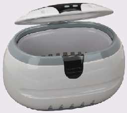
HarborFreight's $30.00 ultrasonic cleaner.

     Further useful tips from DaddyBob and Steve N. of the CS list: For best encapsulation efficiency, blend both the lecithin and your encapsulation solution very well, with a blender, magnetic shaker, or by pure mechanical shaking, until you see no granules or other large debris in solution-then place in the ultrasonic cleaner chamber. At least the use of a common kitchen blender is to be urged for pretreatment of solutions prior to ultrasonic process.

Brooks Bradley writes on this above suggestion:

"First, using some form of blender to enhance/accelerate the process is perfectly acceptable and effective. However, one must understand the limitations of using this modality. To wit: Because the entire encapsulation process is, essentially, a refined homogenization process the researcher is bound within the limits of the chosen process, itself; Using a blender in the early stages of the ultrasonic type protocol, places a limit (especially particle size) on the resultant compounds. As a general rule, the smallest liposomes achievable are going to be larger than 150 nm in size----even after extensive agitating. Therefore, if smaller particles are desired--some procedure must be invoked to achieve this.

Ultrasonic energy is an excellent way to achieve this. Ultrasonic energy applied to solutions having, previously been mixed using mechanical blenders (household type) will improve the encapsulation process greatly (sometimes as much as an order of magnitude) through the immediate size reduction of the encapsulated particle size. Additionally, both power levels and exposure time experienced from the ultrasonic energy have a pronounced effect on the end product: e.g. simply by extending the time exposed to the ultrasonic energy will yield a product with a majority of particles of a markedly reduced physical size (sometimes by more than one-half). Also, by increasing the power spectral density [energy delivered to the target], considerable size and complexity reduction may be achieved (sometimes from larger, multiple-layered liposomes, down to single-layered liposomes of much smaller size). This one characteristic, alone should justify the selection of the larger ultrasonic unit over the smaller one as the larger ultrasonic power level output is much higher. The way to capitalize on this advantage is to limit the depth of the parent solution in the larger ultrasonic unit to 3/4"- 1" deep. Because the distance from the ultrasonic energy source and the mass of the target material DOES, in fact, have a powerful effect on the delivered energy.

Direct visual observation alone will confirm the powerful increase in cavitations (energy field) of the liquid medium. This type of innovation will yield effects that in some cases challenge the results of laboratory-grade, high pressure (over 3000 psi) impact plate systems costing $10,000 and up. What most commercial producers (and labs) do is they RECIRCULATE their candidate solutions in order to achieve smaller and more isolated end products. By extending your exposure time using shallow solutions, do-it-yourselfers can in many cases actually challenge the levels accomplished by these very high dollar commercial machines using their own do-it-yourself homemade systems.

Someone asked the question: does pre-agitation via kitchen blending devices damage or compromise the candidate solutions. The short answer is NO. Almost any type of agitation, aids in the homogenization process."


In regard to particle size and ultrasound cleaner production:


      'We checked for particle size as a major parameter. Second, the power level in watts driving the ultrasonic cleaner transducer and it DOES affect the particle size (at least we found it so). The higher the power, the smaller the majority of the particles (condition held until power levels of our largest lead zirconate-titanate transducers went beyond 1000 watts per transducer.) No effective reduction occurred beyond these power levels (Power Spectral Density evaluations). However, it WAS NOT necessary to reach these power levels to obtain excellent nano-size liposomes. 200 watts driving a transducer at 38K Hertz....yielded excellent results.....even with the high-power lead zirconate units. The larger Harbor Freight unit does, in fact, yield smaller particle liposomes (but the particle size from the small unit was perfectly acceptable for our experiments---and the results gained were, also, quite acceptable as effective in our in vitro evaluations). It should be noted; to get reliable population density numbers, the samples had to be dehydrated completely before viewing with the scanning electron microscope (the same problem is encountered with colloidal silver particle evaluation!). Ultrasonic energy aggitating does facilitate the increased creation of nano-size particles. In fact, Ultrasonic energy was the FIRST energy source to actually achieve this level of size reduction (so I am informed by staff members more conversant with this technology...than am I). Diffraction grating came later.While we have not conducted detailed analyses using calcium ascorbate as the vitamin C component, there seems to be no contravening reason that would seriously modify the excellent results we enjoyed with sodium ascorbate. However, the coefficient of absorption for sodium ascorbate in higher mammals does indicate to be superior to calcium ascorbate (so I am informed). The principal reason calcium carbonate is utilized by most commercial vendors is to mitigate against the alimentary challenges presented to some by the acid form (ascorbic acid). The absorptive ability of sodium ascorbate in humans as against that of ascorbic acid, demonstrates to be over two orders of magnitude ( about 3000 times according to Dr. Gerard Judd). One comment I might add: If a subject is orally consuming LARGE quantities (over 20 grams daily) of vitamin C (especially in the ascorbate form), additional improvement levels, via liposomal additions, in addressing the existing insult may be less than striking (especially if the subject presents with excellent systemic absorption characteristics)....if only because influence levels of the current dosage regimen are reaching near the upper "practical" limits for vitamin C in these cases. However, I have no measured corroboration for such a phenomenon. "

Sincerely, Brooks Bradley.


Several techniques are available for sizing liposomes to a desired size. One sizing method is described in U.S. Pat. No. 4,737,323, incorporated herein by reference. Sonicating a liposome suspension either by bath or probe sonication produces a progressive size reduction down to small unilamellar vesicles less than about 0.05 microns in size. Homogenization is another method that relies on shearing energy to fragment large liposomes into smaller ones. In a typical homogenization procedure, multilamellar vesicles are recirculated through a standard emulsion homogenizer until selected liposome sizes, typically between about 0.1 and 0.5 microns, are observed. The size of the liposomal vesicles may be determined by quasi-electric light scattering (QELS) as described in Bloomfield, Ann. Rev. Biophys. Bioeng., 10:421-450 (1981), incorporated herein by reference. Average liposome diameter may be reduced by sonication of formed liposomes. Intermittent sonication cycles may be alternated with QELS assessment to guide efficient liposome synthesis

First off what is Lecithin? To quote Wikipedia:

" Lecithin is any of a group of yellow-brownish fatty substances occurring in animal and plant tissues and in egg yolk, composed of phosphoric acid, choline, fatty acids, glycerol, glycolipids, triglycerides, and phosholipids. However, lecithin is sometimes used as a synonym for pure phosphatidycholine, a phospholipid that is the major component of its phosphatide fraction. It may be isolated either from egg yolk or from soybeans from which it is extracted chemically (using hexane) or mechanically. It has low solubility in water. In aqueous solution its phospholipids can form either liposomes, bilayer sheets, micelles, or lamellar structures depending on hydration and temperature. This results in a type of surfatant that is usually classified as amphoteric. Lecithin is sold as a food supplement and for medical uses."


What is a liposome? To again quote from Wikipedia:

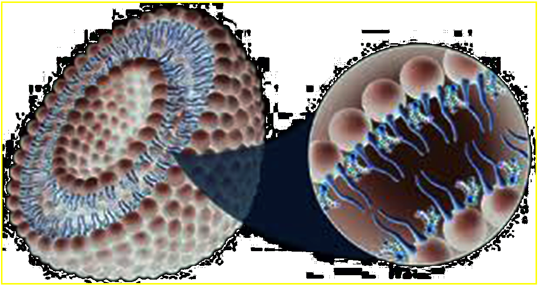
"A liposome is a tiny bubble made out of the same material as a cell membrane. Liposomes can be filled with drugs and used to deliver drugs for cancer and other diseases. Membranes are usually made of phospholipids (lecithin) which are molecules that have a head group and a tail group. The head is attracted to water and the tail which is made of a long hydrocarbon chain is repelled by water. In nature, phospholipids are found in stable membranes composed of two layers (a bilayer). In the presence of water, the heads are attracted to water and line up to form a surface facing the water. The tails are repelled by water and line up to form a surface away from the water. In a cell, one layer of heads faces outside of the cell attracted to the water in the environment. Another layer of heads faces inside the cell attracted by the water inside the cell. The hydrocarbon tails of one layer face the hydrocarbon tails of the other layer and the combined structure forms a bilayer. When membrane phospholipids are disrupted, they can reassemble themselves into tiny spheres, smaller than a normal cell, either as bilayers or monolayers. The bilayer structures are liposomes. The monolayer structures are called micelles. The lipids in the plasma membrane are chiefly phospholipids. Phospholipids are amphiphilic with the hydrocarbon tail of the molecule being hydrophobic; its polar head hydrophilic. As the plasma membrane faces watery solutions on both sides, its phospholipids accommodate this by forming a phospholipid bilayer with the hydrophobic tails facing each other. Liposomes can be composed of naturally-derived phospholipids with mixed lipid chains Liposomes, usually but not by definition, contain a core of aqueous solution; lipid spheres that contain no aqueous material are called micelles, however, reverse micelles can be made to encompass an aqueous environment. Liposomes are used for drug delivery due to their unique properties. A liposome encapsulates a region on aqueous solution inside a hydrophobic membrane; dissolved hydrophilic solutes cannot readily pass through the lipids. Hydrophobic chemicals can be dissolved into the membrane, and in this way liposome can carry both hydrophobic molecules and hydrophilic molecules. To deliver the molecules to sites of action, the lipid bilayer can fuse with other bilayers such as the cell membrane, thus delivering the liposome contents. By making liposomes in a solution of drugs (which would normally be unable to diffuse through the membrane) they can be (indiscriminately) delivered past the lipid bilayer. There are three types of liposomes - MLV (multilamillar vesicles) SUV (Small Unilamellar Vesicles) and LUV (Large Unilamellar Vesicles). These are used to deliver different types of drugs. Liposomes are used as models for artificial cells. Liposomes can also be designed to deliver drugs in other ways. Liposomes that contain low (or high) pH can be constructed such that dissolved aqueous drugs will be charged in solution (i.e., the pH is outside the drug's pI range). As the pH naturally neutralizes within the liposome (protons can pass through some membranes), the drug will also be neutralized, allowing it to freely pass through a membrane. These liposomes work to deliver drug by diffusion rather than by direct cell fusion. Another strategy for liposome drug delivery is to target endocytosis events. Liposomes can be made in a particular size range that makes them viable targets for natura macrophage phagocytosisl. These liposomes may be digested while in the macrophage's phagosome, thus releasing its drug. Liposomes can also be decorated with opsonins and ligands to activate endocytosis in other cell types."

This is an interesting video documenting how a New Zealand farmer with swine flu and later whiteout pneumonia and leukemia came back from the brink of death via Vitamin C in both iv and liposomal forms. Watch:

The renowned Vitmain C researcher and clinician, Dr T. Levy writes:

"For the better part of two years, I actually ignored my own medical observations, since they were in complete conflict with what I felt just had to be true. Also, until the past nine months or so, I had not bothered to educate myself extensively on the body of liposome science that has been accumulating for the past 45 years or so. In a nutshell, I found that liposome encapsulated vitamin C, taken orally, was roughly 10 times more effectively clinically in resolving infectious diseases than the IV-C. Having given thousands of IV-Cs and taken hundreds myself, this was difficult to comprehend, even though the clinical observation was quite straightforward. I subsequently realized that the liposome gave the ultimate bioavailability: intracellular delivery, including the mitochondria, endoplasmic reticulum, and even the nucleus. Furthermore, it was delivered in a non-energy-consuming fashion. IV vitamin C requires an expenditure of energy to eventually reach the intracellular compartment, but liposome encapsulated vitamin C does not. If possible, you do not want to consume energy to get energy-carrying substances inside the cell. It defeats the basic purpose. But let me clear, if it is possible, give a patient both IV-C and oral liposome encapsulated vitamin C. However, if only one is available, the best application is with liposomes orally."

     Liposome made from pure phospholipids (in the absence of cholesterol) will not form at temperatures below the phase transition temperature of phospholipid. If the encapsulated molecule is temperature sensitive (e.g. vitamin C) then pure long chain saturated lipids can not be used in the liposomes because the lipids have to be heated up and the Vit C can not tolerate high temperatures. Phase transition temperature of soy phospholipid is well below room temperature.

     Here is an interesting press release on the use of liposomes to treat lung infections. This may be the key to controling bleeding in racehorses, who probably are infected with pulmonary biofilms:


Monday, March 31, 2008 STUDY CONFIRMS ANTI-INFECTIVE ARIKACE™ EFFECTIVELY PENETRATES MUCUS AND BIOFILM, AND KILLS PSEUDOMONAS, A BACTERIA PLAGUING CYSTIC FIBROSIS PATIENTS

MONMOUTH JUNCTION, NJ, March 31, 2008 Transave Inc. reported today that its lead product candidate, ARIKACE™ (liposomal amikacin for inhalation), may have the ability to penetrate mucus and biofilms, and decrease the number of Pseudomonas aeruginosa lung infections in patients with cystic fibrosis, according to results of a study published in the Journal of Antimicrobial Chemotherapy.

Results of the study titled "Biofilm penetration, triggered release and in vivo activity of inhaled liposomal amikacin in chronic Pseudomanas aeruginosa lung infections," were published in the April issue of the journal. Transave is a biopharmaceutical company focused on the development of next-generation liposomal drug products for inhalation.

ARIKACE is a form of the antibiotic amikacin, which is enclosed in nanocapsules of lipids called liposomes. The study evaluated the ability of the ARIKACE liposomes to pass through patient mucus (sputum) ex vivo and to penetrate the bacteria's biofilm barrier in an established flow-cell in vitro model. The biofilm is a gel-like matrix in the lungs formed by colonies of Pseudomonas that create a protective barrier for bacteria. This prevents patients with cystic fibrosis (CF) from clearing infections even under aggressive antibiotic treatment. It is not practical to observe biofilm interactions in humans; therefore, the Center for Biofilm Engineering at Montana State University has developed a model to microscopically visualize the penetration of liposomes into Pseudomonas biofilms.

Separate studies showed that the small, neutrally charged ARIKACE liposomes facilitated antibiotic passage through the patient mucus layer and penetration into the Pseudomonas biofilm. Prior studies have shown that free aminoglycosides bind to patient mucus and thus have reduced bioactivity in killing Pseudomonas. Using microscopic techniques, the investigators were able to see that Arikace liposomes effectively penetrated and lodged into the spaces within biofilms, where the antibiotic can be released very close to the bacteria.

Another important study enabled investigators to identify bacterial virulence factors secreted by Pseudomonas in the biofilm that trigger release of amikacin from the liposomes. These factors are expected to be concentrated in and near the colonies of Pseudomonas growing in the biofilm within the static mucus of CF lungs. This area is an ideal target for antibiotics and the triggered release of amikacin resulted in what was essentially Pseudomonas suicide.

"Pseudomonas is insidious in the lungs of CF patients who live with chronic infection which is why we developed a liposomal delivery technology small enough for nebulization and able to penetrate the highly-shielded biofilm," said Walter Perkins, Chief Technology Officer at Transave. "Our ultimate goal is to deliver more active antibiotic locally to the site where Pseudomonas resides in the lung, and sustain it there, while reducing the treatment burden with fewer daily doses of drug therapy."

"We know for sure that the liposomes were interacting with, and accumulating in, the biofilm," said Dr. Garth James, Ph.D., director of medical projects at the Center for Biofilm Engineering at Montana State University and a co-author of the study. Montana State University's Center for Biofilm Engineering is the world's largest and oldest biofilm research center.

"You need a drug that can penetrate biofilm in order to kill the infection. This unique drug delivery mechanism may allow that to happen," Dr. James said. "By using an advanced delivery mechanism, you can keep the drug where it needs to be for a longer period of time. A "free" drug inhaled into the lungs would likely be cleared more rapidly."

Excess mucus in the lungs of CF patients is a breeding ground for insidious Pseudomonas bacteria. CF patients with these types of infections are typically treated twice daily for 28 days with an inhaled antibiotic followed by a 28 day drug holiday or "off period." This on/off cycle is often repeated multiple times and patients are surviving longer as a result. However, the commercially available treatments are not designed to penetrate the patient mucus and biofilm, have limited exposure time, and are not able to kill some of the Pseudomonas bacteria protected by the biofilm. The remaining bacteria quickly reproduce during the off treatment period to pretreatment levels and may be more resistant to further antibiotic treatment. This cycle leaves CF patients in a chronic state of infection and can cause further development of hyper-mutated or mucoidal strains of Pseudomonas. Respiratory exacerbations, loss of lung function and death can result.

The study was authored by Drs. Paul Meers and Walter Perkins of Transave Inc., and their colleagues, in collaboration with Garth James, Ph.D., and Steven Fisher, Ph.D., of the Center for Biofilm Engineering at Montana State University. The publication is currently available on the journal's website (jac.oxfordjournals.org/cgi/content/abstract/61/4/859).

The Cystic Fibrosis Foundation provided a $1.7 million award for the clinical program. The Cystic Fibrosis Foundation is the leading organization devoted to curing and controlling cystic fibrosis.

About ARIKACE (liposomal amikacin for inhalation)

ARIKACE is a form of the antibiotic amikacin that is enclosed in nanocapsules of lipid called liposomes. This proprietary next-generation liposomal technology prolongs release of amikacin in the lung while minimizing systemic exposure. The treatment uses biocompatible lipids endogenous to the lung that are formulated into small (0.3 mm) neutrally charged liposomes that enable biofilm penetration and are highly efficient with very low lipid to drug ratio (0.6). ARIKACE can be effectively delivered through nebulization where the small aerosol droplet size (~3.0 mm) facilitates lung distribution. Two Phase II studies are currently being conducted in patients that have CF and Pseudomonas lung infections in Europe and the United States. An abstract on the top line European Phase II data in CF has been accepted for presentation at the 31st European Cystic Fibrosis Conference in Prague in June. ARIKACE has been granted orphan drug status in the United States by the FDA and orphan drug designation in Europe by the European Medicines Agency (EMEA) for the treatment of Pseudomonas infections in patients with CF.

About Transave Inc.

Transave Inc is a biopharmaceutical company focused on the development of innovative, inhaled pharmaceuticals for the site-specific treatment of serious lung diseases. The company's major focus is developing antibiotic therapy delivered via next-generation liposomal technology in areas of high unmet need in respiratory disease. Transave is dedicated to leveraging its advanced liposomal development and commercialization expertise, along with its intellectual property, to bring life extending and enhancing medicines to patients.

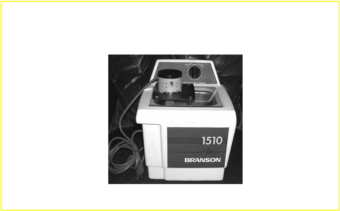
A more advanced ultrasonic unit that I put together to make DIY Lipo-C. A used Branson unit with a plexi-glas lid that supports a electric mixer.